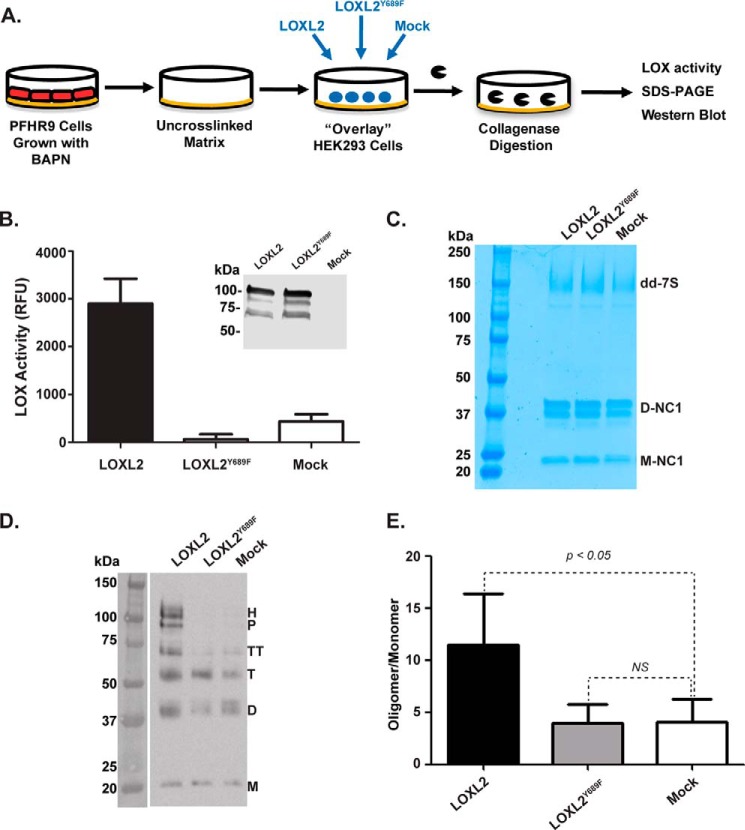
FIGURE 5.
LOXL2 cross-links the 7S dodecamer in a native insoluble basement membrane. A, schematic representation of the overlay assay showing the isolation of PFHR-9 matrix, transient transfection of LOXL2 and LOXL2Y689F of HEK293 cells overlaid on a BAPN-treated substrate matrix, and 7S and NC1 detection after collagenase digestion. B, LOX enzymatic activity in the culture medium of HEK293 cells transfected with either LOXL2 or LOXL2Y689F mutant. Expression of enzymes was confirmed by Western blotting analysis using anti-LOXL2 antibody. Error bars represent S.D. from three independent experiments. C, non-reducing SDS-PAGE analysis of a collagenase digest of PFHR-9 matrices from the overlay assay. 7S-dd, 7S dodecamer; D-NC1, NC1 dimer; M-NC1, NC1 monomer. D, immunoblotting analysis using anti-7S antibody of protein samples from C fractionated under reducing conditions. M, D, T, TT, P, and H indicate the electrophoretic mobility of monomer, dimer, trimer, tetramer, pentamer, and hexamer 7S subunits, respectively. E, quantitation of signal intensities of cross-linked 7S subunits (dimer, trimer, tetramer, pentamer, and hexamer) relative to 7S monomers (uncross-linked) for each lane. Error bars represent S.D. from three independent experiments. Data were analyzed by t test. Statistical significance is shown above the bars. NS, not significant; RFU, relative fluorescence units.

