
FIGURE 1.
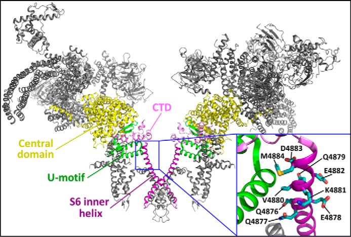
Location of the S6 cytoplasmic region in the three-dimensional structure of RyR2. The 3D structure of two RyR2 monomers (30) is shown. The central domain, U motif, CTD, and S6 inner helix are highlighted. The inset is a close-up view showing the cytoplasmic region of the S6 inner helix encompassing residues 4876QQEQVKEDM4884.