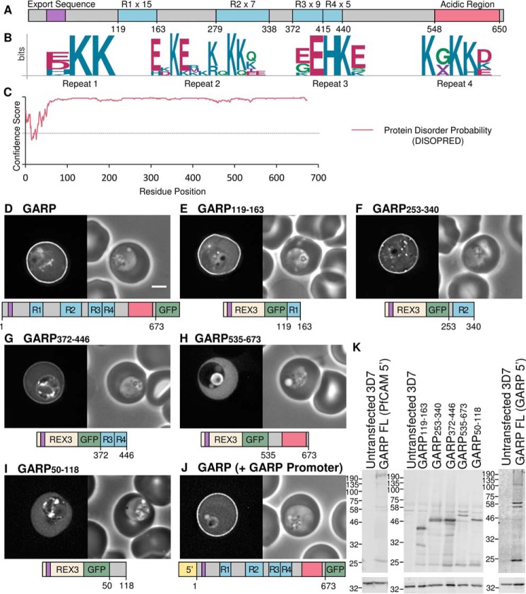
FIGURE 1.
GARP is targeted to the erythrocyte periphery by three lysine-rich repeat regions. A, representation of the GARP protein. The lysine-rich repeating regions are highlighted in blue and labeled R1–R4, and the number of repeating units in each is indicated. The acidic C terminus is shown in red. The export sequence is colored purple and represents both the signal sequence and PEXEL/HT motif. B, sequence logos for the four lysine-rich repeats; residue position is shown on the x axis, and conservation is indicated on the y axis (bits). C, disorder prediction for GARP (using DISOPRED (55)). Amino acids are considered disordered if they have a confidence score >0.5, represented by a dotted line. D–I, GFP-tagged full-length GARP and truncations expressed using the calmodulin promoter in P. falciparum parasites. J, GFP-tagged full-length GARP expressed using the GARP promoter. GFP fluorescence and phase-contrast images are shown on the left and right, respectively. A representation of each construct is shown below. Scale bar, 2 μm. For quantification of fluorescence, see supplemental Fig. 1 and Table 2. K, anti-GFP Western blot (top), with anti-HAP used to confirm equal loading (bottom).

