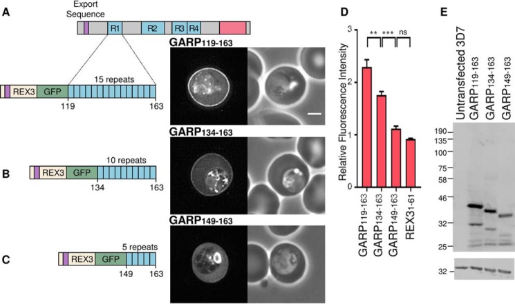
FIGURE 2.
Truncating the first charged repeat of GARP decreases targeting efficiency. A–C, truncated fragments of the first lysine-rich repeat sequence (blue) of GARP, GFP-tagged and expressed in P. falciparum parasites. Expressed proteins are shown on the left, with dotted lines indicating the region cloned from the full-length protein. The export sequence (purple) of REX3 was used to drive export of each fragment. GFP fluorescence and phase-contrast images are shown on the left and right, respectively. Scale bar, 2 μm. D, the ratio of the fluorescence intensity adjacent to the erythrocyte membrane relative to the erythrocyte cytoplasm for the indicated proteins is shown. Error bars, S.E. ns, *, **, ***, and ****, not significant (p > 0.05), p ≤ 0.05, p ≤ 0.01, p ≤ 0.001, and p ≤ 0.0001, respectively. E, anti-GFP Western blot (top), with anti-HAP used to confirm equal loading (bottom).

