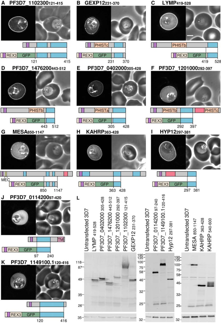
FIGURE 4.
Multiple lysine-rich repeating sequences from P. falciparum proteins target the erythrocyte periphery. A–K, identities of proteins are shown above each image. Shown are GFP localization (left) and a phase-contrast image (right). A representation of the full-length protein is shown below each image, with the lysine-rich repeat regions in blue, export sequences (signal sequence and PEXEL/HT motif) in purple, acidic sequences in red, PRESAN/PHIST domains in orange, predicted transmembrane domains in pink, and the MESA erythrocyte cytoskeleton-binding (MEC) motif in yellow. Dotted lines, protein sequence cloned into each GFP-tagged construct, shown below the full-length protein. Protein schematics are approximately to scale, with MESA downscaled by one-half. Scale bar, 2 μm. L, anti-GFP Western blots of all parasite lines (top). Anti-HAP was used to confirm equal loading (bottom).

