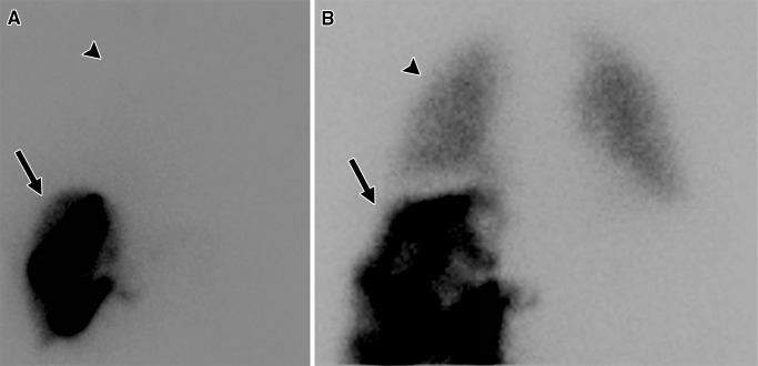
Figure 1:
Examples of 99mTc-MAA scintigraphic images obtained during evaluation prior to radioembolization. After 99mTc-MAA was injected into the hepatic artery to be treated, the patient underwent planar scintigraphy or SPECT to calculate LSF. A, In a patient with low LSF (4.4%), activity was primarily found in the liver (arrow), with little activity detected in the lungs (arrowhead). B, In a patient with high LSF (16%), significant activity was detected in the lungs (arrowhead), which was compared with the activity in the liver (arrow) to calculate LSF.

