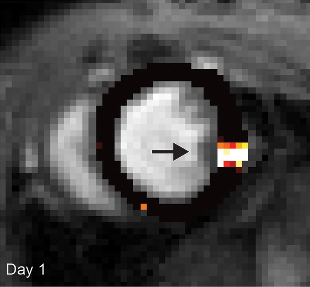
Figure 1a:

(a, b) Longitudinal cardiac CEST images of the survival of Eu-HP-DO3A–labeled cells in C3H mice. Twenty-four hours after implantation (a), significantly increased MTRasym values were observed adjacent to the inferior papillary muscle (arrow), corresponding to the location of Eu-HP-DO3A–labeled cells. After 20 days (b), increased MTRasym was still observed in the same myocardial region surrounding the inferior papillary muscle (arrow). The proliferation of labeled cells and the likely dilution of Eu-HP-DO3A with cell division reduced the MTRasym values of the graft relative to day 1. (c) Photomicrograph (hematoxylin-eosin stain; original magnification, ×4) of the corresponding histologic slice demonstrates a graft of proliferating cells (blue) adjacent to the inferior papillary muscle in a similar location to increased MTRasym values seen on b. (d) Higher magnification photomicrograph (hematoxylin-eosin stain; original magnification, ×20) of the area on c enclosed within the black box demonstrates the presence of proliferating cells (arrows) near the endocardial surface at the boundary of the papillary muscle.
