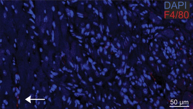
Figure 5b:
Photomicrographs show macrophage staining in C3H cardiac tissue. (a) Nuclear staining (4ʹ6-diamidino-2-phenylindole·2HCl, or DAPI; original magnification, ×40 composite) of a cross-section of cardiac tissue from a mouse that underwent implantation of Eu-HP-DO3A–labeled C2C12 cells. (b) Photomicrograph with higher magnification of a region of interest within the cell graft (original magnification, ×400 composite) is shown after immunostaining for murine macrophages (F4/80, red) and cell nuclei (DAPI, blue). Murine macrophages (arrow) were not present in high numbers in C3H cardiac tissue 20 days after cell implantation. Corresponding images from a C57BL/6J mouse are found in Figure E4 (online).

