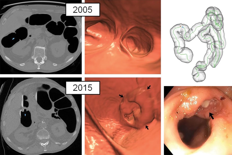
Figure 4:
Normal initial CT colonography screening (even in retrospect) in asymptomatic man (60 years old at initial screening; 70 years old at repeat screening) with obvious cancer detected 10 years later at follow-up screening. Top: on two-dimensional (2D) (left) and three-dimensional (3D) (middle) images from the initial CT colonography screening in 2005, no focal lesions were identified, even in retrospect, in the area of subsequent cancer in 2015. Bottom: 2D (left) and 3D (middle) images from repeat CT colonography screening in 2015 now show a semiannular mass near the hepatic flexure (arrows). Initial biopsy of mass (arrow) from same-day colonoscopy (right) revealed only tubular adenoma, but repeat colonoscopy with biopsy 4 months later revealed adenocarcinoma at pathologic evaluation.

