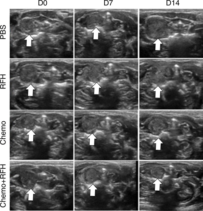
Figure 5b:
(a) Optical and x-ray images and (b) US images show tumor response as assessed according to signal intensity (yellow-red colors on a) and tumor size (arrows on b) in the four groups. There was a larger decrease in both signal intensity and tumor size in the combination therapy group compared with the other three groups. Graphs show quantitative analysis, which further confirms the significant decrease in both photon intensity according to (c) optical imaging and (d) relative tumor volume according to US imaging after combination therapy (chemotherapy and RF hyperthermia) compared with the other groups. (e) Photographs of representative tumors harvested from four different animal groups show smallest tumor (arrows) size in the combination therapy group compared with the others (left and middle columns, arrowheads indicate esophageal wall). Photomicrographs of apoptosis analysis further confirmed more apoptotic cells (blue dots) in the combination therapy group than in the other three groups (right column, magnification, ×20). RFH = RF heat.

