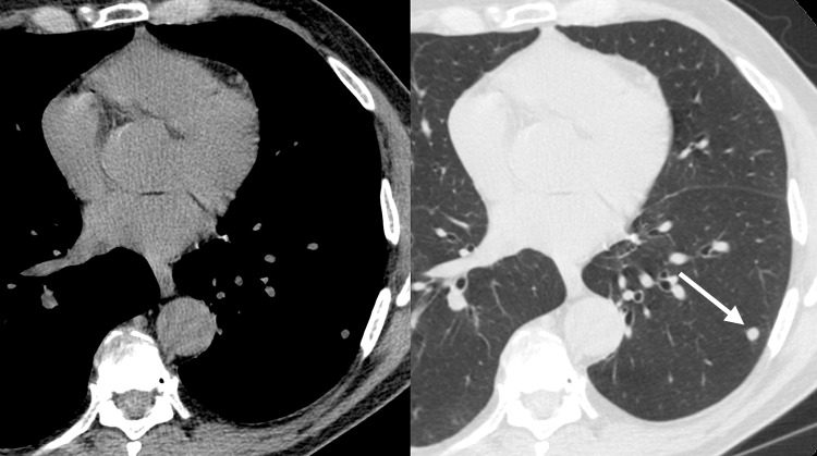
Figure 3e:
Improved nodule visibility with tomosynthesis. A 7-mm lower-left-lobe CT-confirmed nodule was not visible on the (a) conventional chest radiograph, (b) DE tissue image, or (c) DE bone image but was visible on the (d) tomosynthesis image (arrow). (e) Axial CT images obtained with two different window settings (mediastinal window and lung window) show the nodule (arrow on the right image).

