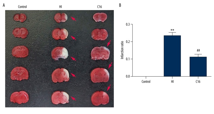
Figure 3.
The effects of C16 administration on brain tissue loss at 24 h after hypoxia-ischemia. (A) Representative samples of TTC-stained coronal sections were obtained 24 h after hypoxia-ischemia. Notable cerebral infarction region was observed in the HI group. (B) After HI, the infarct ratio was increased dramatically, and the infarct ratio was clearly lower after intraperitoneal C16 administration. ** P<0.01 vs. Control group, ## P<0.01 vs. HI group.

