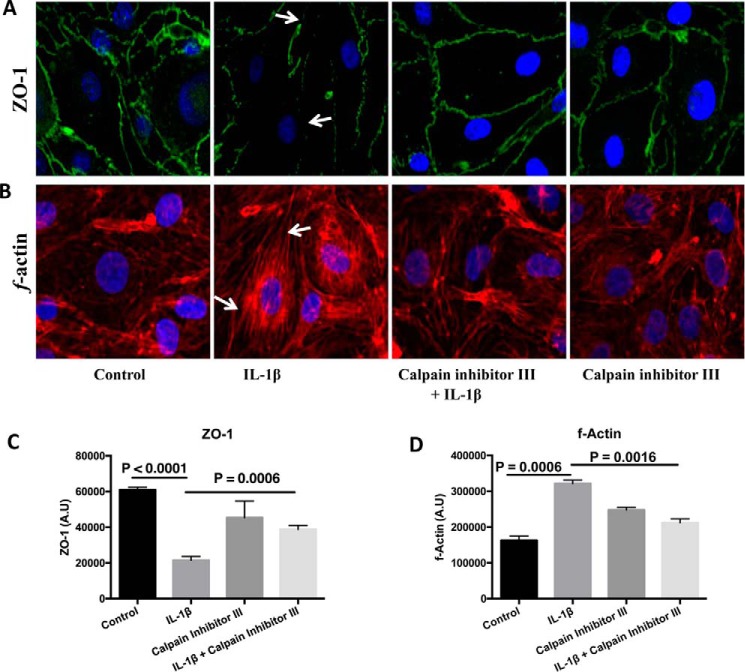
FIGURE 4.
IL-1β treatment-induced ZO-1 junctional disruption and F-actin stress fiber formation is reduced by pretreatment with calpain inhibitor III (A–D). IL-1β treatment-induced ZO-1 junctional disruption and F-actin stress fiber formation are shown by white arrows in A and B, respectively. ZO-1 junctional integrity and F-actin stress fiber formation were assessed using immunofluorescence localization and rhodamine phalloidin techniques, respectively (n = 4 for each study). The changes in ZO-1 localization and the formation of F-actin stress fibers were further determined using ImageJ software and presented as arbitrary units (C and D, respectively; p < 0.05). Under normal physiological conditions actin filaments are randomly distributed throughout the cell, but agents that induce endothelial hyperpermeability induce them to reorganize themselves into stress fibers that are linear parallel bundles across the cell interior (following IL-1β treatment as pointed by the arrows in B) and also exhibit increased binding to rhodamine phalloidin (as shown in B and D).

