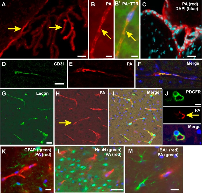
FIGURE 3.
The presence of PA in the choroid plexus and vascular endothelial cells. Immunofluorescence staining with a monoclonal antibody to PA under a confocal microscope showed a clear PA positivity in the choroid plexus in the cerebral ventricles (red color; arrows in A and B). Double immunofluorescence staining of PA (B; red color) with TTR (B′; green color) showed PA co-localization with TTR (yellow color in merged image in B′). Double immunostaining of PA (red color in C, E, F, H, I, J, K, and L) with DAPI (C; blue color), CD31 (D; green color), lectin (G; green color), PDGFRβ (J; green color), GFAP (K; green color), NeuN (L; green color), and microglia (M; red color) was also performed to show co-localization. PA immunostaining was co-localized with CD31 (F; yellow color) and lectin staining (I; yellow color), indicating that PA is expressed by endothelial cells. PA did not show co-localization with pericytes (J; bottom panel), which surround the microvessels, or with astrocytes (K), microglia (M), and neurons (L). Scale bars, 50 μm.

