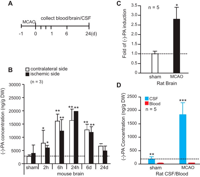
FIGURE 4.
Cerebral ischemia elevates the level of (−)-PA in the CSF and brain. A, schematic diagram of MCAO model. The middle cerebral artery was occluded for 1 h and followed by up to 24 days of reperfusion. Blood and brain tissues were collected at 2 h, 6 h, 24 h, 6 days, and 24 days after reperfusion. CSF was collected after 24 h of reperfusion. Samples were subjected to UPLC/MS/MS analysis. B shows the elevated (−)-PA level in both the ipsilateral and contralateral sides of the ischemic brain. Rat brains were collected after 24 h of reperfusion, and -fold induction of (−)-PA was determined and is presented in C. CSF and blood were taken after 24 h of reperfusion from MCAO rats, and the concentration changes are shown in D. Error bars represent the mean ± S.D. * indicates p < 0.05, ** indicates p < 0.01, and *** indicates p < 0.001 (one-way ANOVA with Tukey's post hoc analysis; n = 5). d, days.

