FIGURE 4.
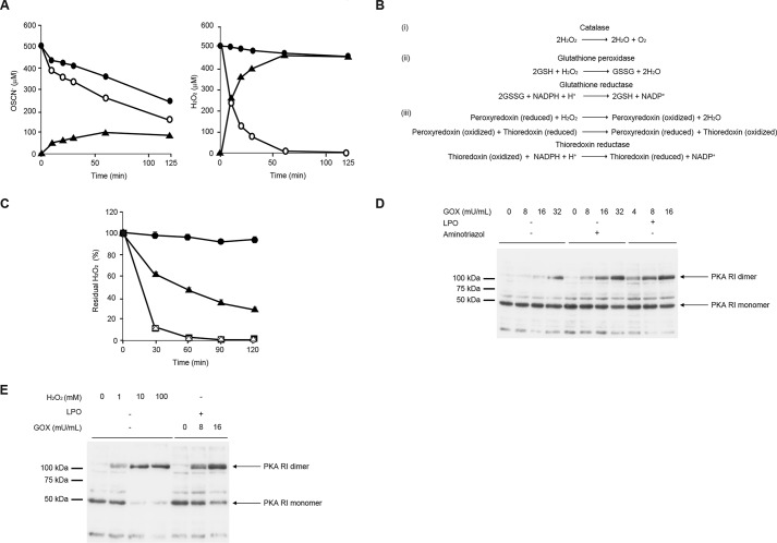
H2O2 is detoxified mainly by catalase in airway epithelial cells. A, medium was prepared with 11 mm β-d-glucose, 3 mm HSCN, 16 milliunits/ml GOX in the presence (left) or absence (right) of 10 μg/ml LPO. The medium was incubated with H292 cells (open circle) or without H292 cells (closed circle) for 5 h. Produced OSCN− (left) and H2O2 (right) are depicted. The consumed amounts (closed triangle) were calculated by subtracting the concentrations in the presence of H292 cells from the concentrations in the absence of H292 cells. B, chemical reactions of three H2O2 detoxification systems. C, H292 cells were incubated for 5 h with 100 mm aminotriazole (closed triangle) or 1 mm BCNU (open triangle) or 1 μm auranofin (closed square) or no inhibitor (open circle) using the medium prepared in A. The medium alone, without the cells, is depicted as a closed circle. The residual H2O2 ratios compared with the initial amounts at the indicated times are depicted. D, H292 cells were stimulated for 5 h with medium containing 11 mm β-d-glucose and 3 mm HSCN with the indicated concentrations of GOX in the presence or the absence of 10 μg/ml LPO and/or 100 mm aminotriazole. E, H292 cells were stimulated with medium containing the indicated concentrations of H2O2 alone or 11 mm β-d-glucose, 3 mm HSCN, and 10 μg/ml LPO with the indicated concentrations of GOX for 5 h. D and E, cell lysates were applied to Western blotting under non-reducing conditions using anti-PKA antibodies. All experiments were performed at least more than two times.

