
FIGURE 5.
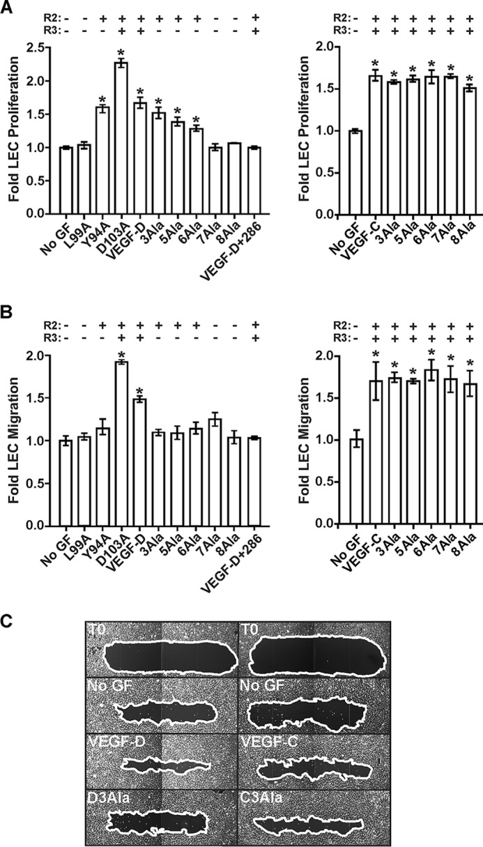
Analyses of the role of N-terminal α-helices of mature VEGF-D and VEGF-C for proliferation and migration by LECs. A, LEC proliferation assays. Adult LECs were treated with VEGF-DΔNΔC (VEGF-D), VEGF-CΔNΔC (VEGF-C), or their variants or left untreated (No GF). VEGF-D+286, combination of VEGF-DΔNΔC and a 10-fold molar excess of mAb 286. y axes represent proliferation by LECs stimulated with growth factor relative to that of unstimulated cells. x axes denote VEGF-D variants (left) and VEGF-C variants (right) used in assays. B, LEC migration assay. The capacity of variant proteins to induce cell migration was assessed in a scratch wound assay. Neonatal LECs were wounded, and the amount of wound closure was calculated for each variant as described under “Experimental Procedures.” y axes show migration of cells stimulated with growth factor relative to that of unstimulated cells. x axes denote VEGF-D variants (left) and VEGF-C variants (right) used in assays. C, images of selected scratch wounds. Wounds were imaged immediately post-wounding (T0, two examples) and after 24-h treatment with VEGF-DΔNΔC, VEGF-CΔNΔC, or the 3Ala variant of each (D3Ala and C3Ala, respectively). No GF, two results after 24 h with no growth factor. White lines, edges of the wounds. In A and B, the capacity of variants to activate VEGFR-2 (R2) or VEGFR-3 (R3) is indicated above the graphs, and asterisks indicate that results differ from No GF in a statistically significant fashion, as assessed by one-way analysis of variance with Tukey's post hoc test. The amounts of VEGF-D or VEGF-C variants were matched in each assay.