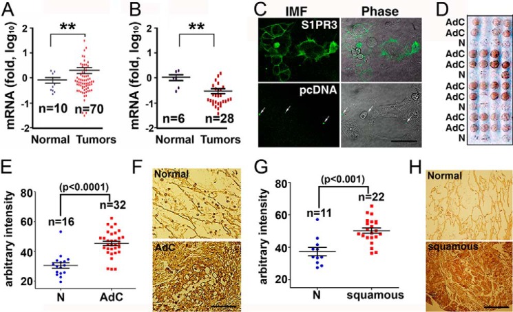
FIGURE 1.
Up-regulation of S1PR3 in human lung adenocarcinomas. A, qPCR quantitation of S1PR3 mRNA in cDNA arrays of human lung adenocarcinoma specimens (OriGene, HLRT101 and HLRT105). **, p < 0.01, Student's t test. B, qPCR quantitation of S1PR2 mRNA in a cDNA array of human lung cancers (OriGene, HLRT105). **, p < 0.01, Student's t test. C, HEK293 cells were transfected with S1PR3 or pcDNA vector. Transfected cells were immunostained with anti-S1PR3 (Cayman Chemical) (IMF, left panels). Arrows, nonspecific fluorescent precipitates used for image orientation. Scale bar = 33 μm. D, anti-S1PR3 staining of human lung adenocarcinoma tumor microarray (Accumax 306). AdC, adenocarcinoma; N, adjacent normal lung tissue. E, immunostaining intensity was quantitated with the National Institutes of Health ImageJ software. Data, analyzed with GraphPad Prism 5 software, are shown as mean ± S.E. Statistical significance was analyzed by Student's t test. F, representative images of anti-S1PR3 staining of human lung adenocarcinoma and the respective adjacent normal lung epithelial tissue. G, quantitation of anti-S1PR3 staining of human lung squamous carcinoma microarray (Accumax 306). Data are mean ± S.E. Statistical significance was analyzed by Student's t test. H, representative images of anti-S1PR3 staining of human lung squamous carcinoma and the respective adjacent normal lung epithelial tissue.

