FIGURE 2.
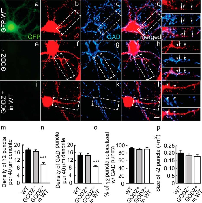
Deficits in GABAAR postsynaptic clustering and inhibitory synapse density in GODZ KO cortical neurons co-cultured with WT neurons. GFP-transgenic WT cultures and GODZ KO cortical neurons were cultured as pure cultures, or the mutant neurons were co-cultured 1:9 with an excess of GFP-expressing WT neurons. GFP autofluorescence was used to distinguish WT from mutant neurons. a–l, representative micrographs of a GFP-WT neuron (a–d; note the GFP autofluorescence in a (green)), a GODZ KO neuron (e–h), and a GODZ KO neuron co-cultured with WT neurons (i–l) immunostained for the γ2 subunit (b, f, and j; red) and GAD (c, g, and k; blue) with corresponding merged images shown in d, h, and l. Boxed dendritic segments are shown enlarged in the right-most column of micrographs with arrows pointing to colocalized pre- and postsynaptic structures of axons that have grown along and seem to adhere to dendrites and arrowheads pointing to axons (blue) that have grown across dendrites with minimal contact. m–p, summary quantifications of the density of γ2 (m) and GAD (n) immunoreactive puncta, their colocalization (o), and the size of γ2 puncta (p). Note the drastic reduction in the density of both pre- and postsynaptic structures selectively in GODZ KO neurons that were grown in the presence of an excess of WT neurons, whereas the density of puncta in pure KO cultures remained unaffected. In mutant neurons grown in competition with WT neurons, the defect in synaptic localization of GABAARs resulted in loss of innervation by WT neurons. Bar graphs with error bars indicate means ± S.E. ***, p < 0.001, ANOVA, Tukey tests. Scale bar, 5 μm.

