FIGURE 7.
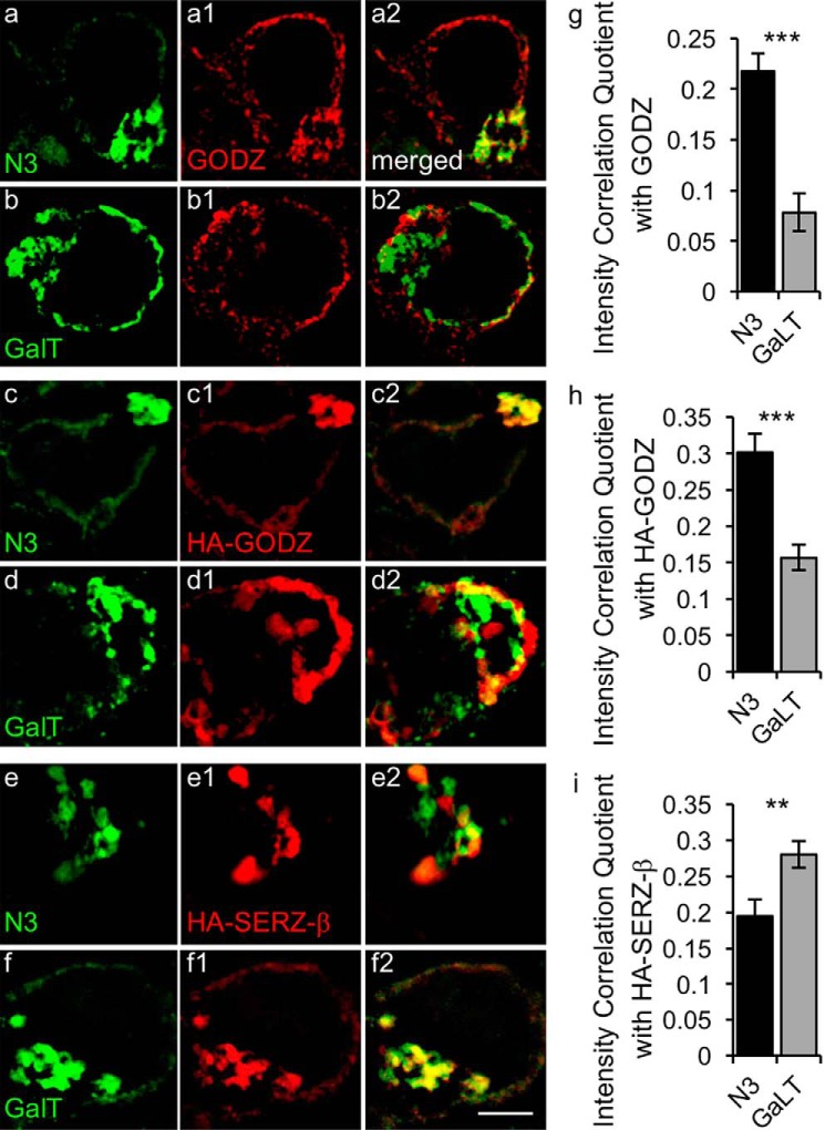
Differential enrichment of GODZ and SERZ-β in cis versus trans compartment of the Golgi complex. a–f, representative SIM images of HEK293T cells transfected with GFP-N3 (a, c, and e) or GalT-YFP (b, d, and f) together with HA-GODZ (c and d) or HA-SERZ-β (e and f). Cells were immunostained for GFP- or YFP-labeled Golgi markers (a–f; green) and either endogenous GODZ (a1 and b1; red) or transfected HA-GODZ (c1 and d1; red) or transfected HA-SERZ-β (e1 and f1; red). Colocalization of the Golgi markers with GODZ or SERZ-β is evident in the merged images depicted in the right-most column (a2–f2; yellow). g–i, summary quantification of colocalization by intensity correlation analyses of immunofluorescence in the Golgi area for the indicated Golgi markers with endogenous GODZ (g), transfected HA-GODZ (h), and transfected HA-SERZ-β (i). Note that both endogenous GODZ and transfected HA-GODZ were strongly colocalized with the cis-Golgi marker GFP-N3 but not with the trans-Golgi marker GalT-YFP. By contrast, HA-SERZ-β showed greater colocalization with GalT-YFP than with GFP-N3. Bar graphs with error bars indicate means ± S.E. **, p < 0.01; ***, p < 0.001, t tests. Scale bar, 2.5 μm.

