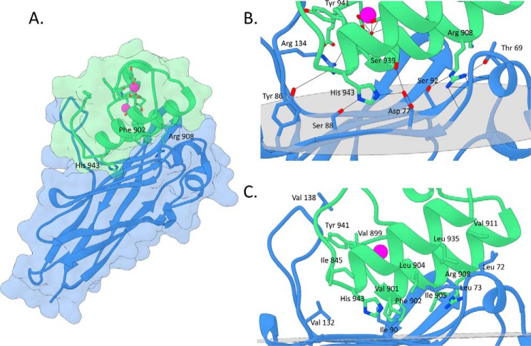
FIGURE 2.
Structure and cohesin-dockerin interface in the R. flavefaciens CohScaC-Doc3 complex. A, structure of CohScaC-Doc3 complex with the dockerin in green and the cohesin in blue. Phe-902, Arg-908, and His-943 that dominate cohesin recognition are labeled and shown as stick configuration. Ca2+ ions are depicted as purple spheres. B, polar interactions at the complex interface. C, hydrophobic interactions at the complex interface. The most important residues in both types of interaction are shown as sticks. The transparent gray disk in B and C marks the plane defined by the 8-3-6-5 β-sheet, where the β-strands form a distinctive dockerin interacting plateau.

