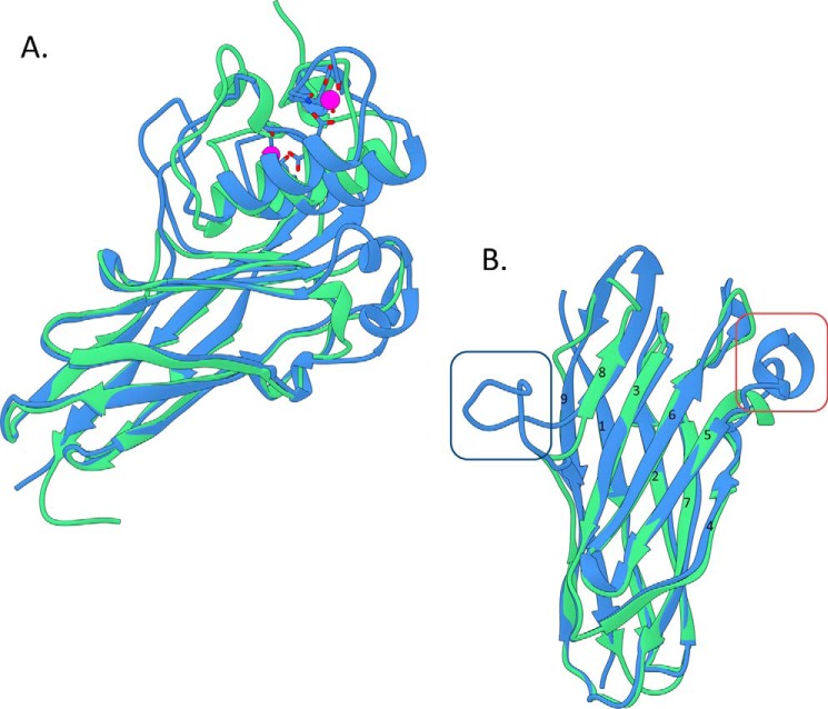
FIGURE 3.
Overlay of the R. flavefaciens CohScaC-Doc3 complex with the A. cellulolyticus type I cohesin-dockerin complex. A, overlay of CohScaC-Doc3 (depicted in blue) with the AcScaCCoh3-ScaBDoc type I complex from A. cellulolyticus (depicted in green, PDB code 4UYP), with the dockerins rotated 180° relative to each other, showing the high degree of overall similarity. B, overlay of both cohesins isolated from the complexes and rotated ∼90° down and right relatively to A, with the dockerin interacting plateau in the first plane. This view highlights the main differences between the two cohesins that consist of the large β-flap extension that interrupts β-strand 8 (dark blue box) and the well defined α-helix connecting β-strands 4 and 5 (red box). These two structural elements together with the loop formed by the distal part of β-strand 8 and the proximal section of β-strand 9 that is tilted toward the dockerin form a claw-like interaction interface.

