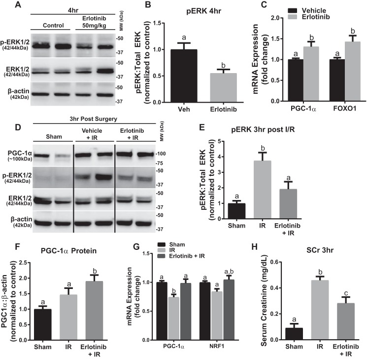
FIGURE 7.
Erlotinib blocks ERK1/2 phosphorylation in naïve mice and following IR, preventing decreases in PGC-1α and NRF1 expression. A, representative immunoblot of phosphorylated ERK1/2 after 4 h of treatment with erlotinib in mouse kidney cortex. p, phosphorylated; MW, molecular weight. B, densitometry analysis of phosphorylated ERK1/2 when compared with total ERK1/2 following 4 h of erlotinib treatment. C, mRNA expression of PGC-1α and FOXO1 at 4 h in mouse cortex following erlotinib treatment. D, representative immunoblot of phosphorylated ERK1/2 and total ERK1/2, as well as PGC-1α following IR AKI. E, densitometry analysis of phosphorylated ERK1/2 when compared with total ERK1/2 following 3 h of IR AKI. F, densitometry analysis of PGC-1α protein when compared with β-actin following 3 h of IR AKI. G, mRNA expression of PGC-1α and NRF1 following 3 h of IR AKI. H, serum creatinine was assessed 3 h after IR AKI. Data are represented as mean ± S.E., n ≥ 4. Different superscripts indicate statistically significant differences (p < 0.05).

