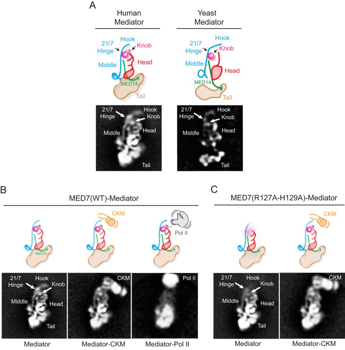
FIGURE 6.
EM analysis of wild-type and MED7(R127A/H129A) human Mediators. A, a 2D class average obtained from stained particle images of wild-type human Mediator and a corresponding diagram summarizing the organization of the head, middle, and tail modules, the hook, and MED14 (left), based on comparison with wild-type yeast mediator (right). B, 2D class averages obtained after alignment and clustering of stained particle images from a wild-type human Mediator preparation. Free Mediator (left) and Mediator associated with kinase module (Mediator-CKM, center) or Pol II (Mediator-Pol II, right) were observed. The head, middle, and tail modules and the MED19-containing “hook” region at top of the middle module can be readily identified. The arrowhead marks the approximate position of the MED21-MED7 hinge region and the MED7(R127A-H129A) mutation, also inferred by comparison with the yeast Mediator structure (6). The Mediator-Pol II average is blurred by variability in the relative position of Mediator and Pol II, exacerbated by disruption of the complex after preservation in stain. C, class averages of free (left) and CKM-associated MED7(R127A/H129A) human Mediator. As illustrated in the diagrams, the MED7(R127A/H129A) mutation increases mobility of the hook and knob portions of the Middle module.

