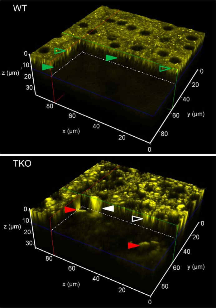
FIGURE 1.

Differences between WT and TKO mice in the distribution of fluorophores in the RPE and photoreceptor cells. TPM 3D reconstructions of the RPE/retina interface in 3-month-old WT and Mertk−/−Abca4−/−Rdh8−/− (TKO) mice are shown. The center of the RPE was set at z = 0 μm with the photoreceptor space starting at 8 μm below. In TKO mice, red arrowheads point to large (over 20 μm) fluorescent granules that extend from z ≈ 6 μm below the RPE cell layer (17). Fluorescent granules of this size, shape, and location that display an emission spectrum with a broad maximum at 600 nm were previously identified as infiltrating microglia/macrophages (17). Another type of large fluorescent granule is indicated with a white arrowhead. These granules extended throughout the thickness of the RPE and into the space below. Smaller granules in the RPE are indicated with a white-outlined black arrowhead. In WT mice, fluorescence in the photoreceptor space was faint and uniform. Most fluorescent structures in the RPE, indicated with green outlined transparent arrowheads, represent retinosomes (retinyl ester-containing lipid droplets) (18, 22) characterized by their 1-μm size and emission spectra with a broad maximum at 500 nm. Microvilli visible at about 5 μm below the center of RPE are indicated with solid green arrowheads.
