
FIGURE 5.
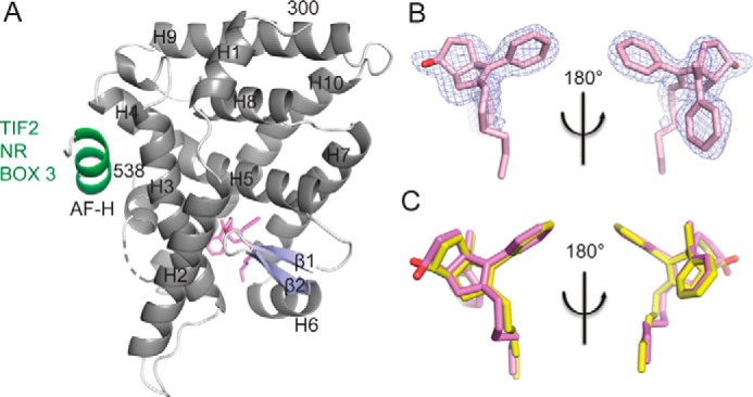
Crystal structure of LRH-1 bound to an RJW100 isomer with endo stereochemistry. A, overall structure of the LRH-1 ligand binding domain with SR-RJW100 (violet) and a peptide derived from the Tif2 co-activator (green). Dashed line, region of disorder in the protein backbone that could not be modeled. B, omit map (FO − FC, contoured at 2.5 σ) showing that a single enantiomer is bound in the structure. C, superposition of SR-RJW100 with RR-RJW100 (yellow) showing a very similar position in the binding pocket.