
FIGURE 6.
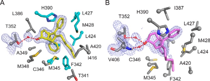
Residues interacting with LRH-1 agonists. Close-up views of the binding pockets from the structures of LRH-1 bound to RR-RJW100 (A) or SR-RJW100 (B), depicting side chains of amino acid residues that interact with each ligand. A, residues that also interact with GSK8470 are shown in cyan, whereas unique interactions made by RR-RJW100 are shown in gray. Portions of the electron density maps are shown to highlight the interactions with Thr-352 through water (FO − FC, contoured to 1σ).