
FIGURE 7.
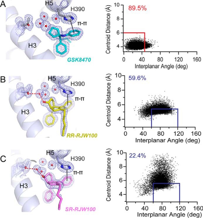
π-π-stacking with residue His-390 differs among agonists. A–C, left, views of the different types of π-π-stacking utilized by GSK8470 (A), RR-RJW100 (B), and SR-RJW100 (C). Right, MDS monitoring the distances between ring centroids (x axis) and angle between the ring planes (y axis) for the ligand phenyl group and His-390 at each time increment of the 200-ns MDS. The red and blue boxes indicate when face-to-face and edge-to-face π-π-stacking occurred, respectively. The numbers in the top left corners indicate the percentage of time spent π-π-stacking during the MDS.