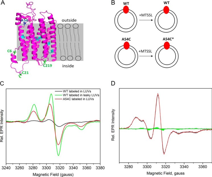
FIGURE 2.
EPR measurements of MTSSL-labeled GtACR1-inserted LUVs to detect the insertion orientation. A, in the predicted structure of GtACR1, the six cysteines in cyan are in the transmembrane helices, and the three cysteines in green face the extracellular side; Ala-54 shown in blue faces the cytoplasmic side. The labels inside and outside refer to the LUVs, of which the lipid bilayer is displayed in gray. B, GtACR1_WT-inserted LUVs (top) and GtACR1_A54C-inserted LUVs (bottom) were incubated with MTSSL. Ala-54 is predicted to face the cytoplasmic side of GtACR1 (panel A), and inserted A54C is labeled by MTSSL. The protein is colored in red, LUVs are represented as two concentric circles, and the spin label is indicated as an asterisk. C, EPR spectra of the frozen LUVs from different treatments were normalized based on their protein concentrations as described under “Experimental Procedures.” The EPR signals are shown for: labeled WT-inserted LUVs, black; labeled WT inserted in leaky LUVs, green; and labeled A54C-inserted LUVs, red. Rel., relative. D, room temperature continuous wave EPR spectra of GtACR1_A54C-inserted LUVs in the dark and light-activated states. The black and red lines are the dark and light spectra, respectively, and the green areas are the light-minus-dark difference spectrum.

