FIGURE 3.
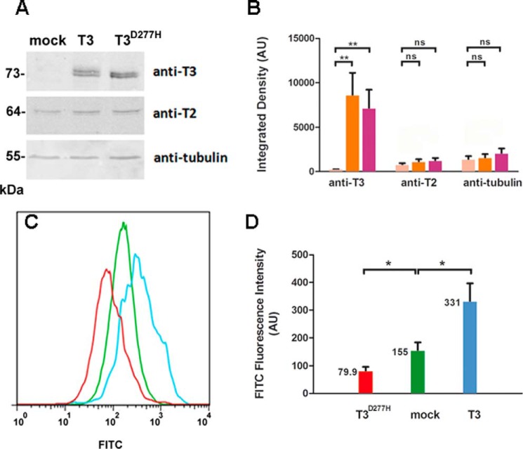
Effect of T3lec on O-GalNAc glycosylation in vivo. CHO ldlD cells were transfected with mock vector, ppGalNAc-T3 (T3), or catalytically inactive mutant ppGalNAc-T3D277H (T3D277H). A, overexpression of recombinant proteins was assayed by Western blotting using anti-ppGalNAc-T3 antibody with anti-ppGalNAc-T2 (T2) and anti-tubulin antibodies as internal control and loading control, respectively. B, bands of Western blotting by using anti-ppGalNAc-T3, anti-ppGalNAc-T2, and anti-tubulin antibodies on CHO ldlD cells transfected with mock vector (pink), ppGalNAc-T3 (orange), or ppGalNAc-T3D277H (fuchsia) were quantified by using ImageJ software. Error bars represent S.D. of three independent experiments. C, effects of mock vector (green), ppGalNAc-T3 (light blue), and ppGalNAc-T3D277H (red) overexpression on O-GalNAc glycosylation were evaluated by flow cytometry of cells stained with biotinylated VVL. D, fluorescence intensity values of FITC (expressed in arbitrary units (AU)) corresponding to means and S.D. of three independent experiments. **, p < 0.01; *, p < 0.05; ns, not significant.

