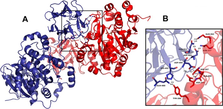
FIGURE 7.
Interaction between catalytic and lectin domains in crystal structure of ppGalNAc-T2 (Protein Data Bank code 2FFV). The two ppGalNAc-T2 molecules of the asymmetric unit exhibit conserved interaction regions (A). The enlarged view shows the amino acids corresponding to the lectin domain (blue) and catalytic domain (red) involved in the interaction (B). Hydrogen bonds are represented with gray dashed lines, and atomic distances are in Å. The figure was prepared using PyMOL.

