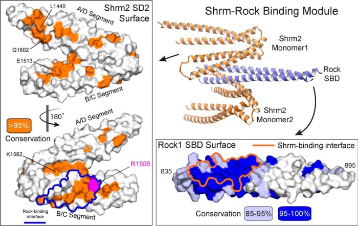
FIGURE 3.
The conserved SD2-SBD interface. Conservation from a multiple sequence alignment of 23 Shrm or 33 Rock1 proteins was mapped onto a surface representation of each protein. Residues with over 95% identity within this alignment are colored orange for Shrm (left box), and residues over 85% identical (light blue) and over 95% identical (blue) are indicated on the Rock1 surface (lower right box). The boundaries of the observed binding interface are outlines for both proteins. A ribbon diagram of the complex is shown for reference.

