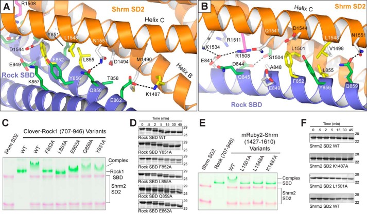
FIGURE 4.
The molecular basis for Shroom-Rock recognition. A and B, ribbon diagram of the Shrm-Rock interface featuring important residues as sticks. Residues previously shown to mediate Shrm-Rock interactions are indicated in green, additional residues shown to mediate interactions as a part of this study are colored yellow, and the position Arg-1508 shown to mediate neural tube defects in mice is shown in magenta. Selected hydrogen bonding interactions within the interface are indicated with black dotted lines. C and E, mutants in the Shrm-Rock interface block complex formation. Wild-type or mutant Shrm SD2 (mRuby-SD2) or Rock1 SBD (residues 707–946 fused to Clover) were mixed as indicated, and their ability to form a complex was assayed by native PAGE, imaged at 460 or 630 nm, and presented as a false colored overlay. D, WT and mutant Rock1 (residues 707–946) proteins were subjected to limited proteolysis using 0.025% trypsin, and samples were taken at the indicated time points. In this assay, the mutant proteins behaved similarly to wild type. F, limited proteolysis for Shrm SD2 (residues 1427–1610) mutants was performed as in D.

