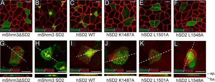
FIGURE 5.
Shrm SD2 and Rock SBD point mutations disrupt binding in vitro. A–F, MDCK cells were transiently transfected on Transwell filters with the indicated constructs and stained to detect Shrm (green) and ZO-1 (red). Scale bars represent 10 μm. Images are projections of 0.5-μm confocal sections. G–L, MDCK cells were transiently co-transfected on Transwell filters with Myc-Rock (residues 681–942) and the indicated constructs and then stained to detect Shrm (green) or Myc (red). Scale bars represent 10 μm. Images are projections of 0.5-μm confocal sections. ap., apical surface; ba., basolateral surface; hSD2, human SD2.

