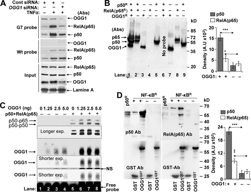
FIGURE 3.
OGG1 facilitates NF-κB binding to its motif. A, OGG1 and NF-κB's occupancy of 8-oxoG-containing DNA in NE. NE (50 μg per sample) from ±TNFα-exposed, OGG1-expressing, and depleted cells were incubated with G7 (upper panel) or WT probe (middle panel) for 5 min, and protein DNA complexes were pulled down using magnetic streptavidin beads. The washed pellets were subjected to SDS-PAGE and immunoblotting using Abs to OGG1, p50, or RelA(p65). Lower panel (input): NE (3 μg per sample) was subjected SDS-PAGE and immunoblotted by using Ab to OGG1 and lamin A. B, OGG1 increases DNA occupancy of NF-κB. Annealed RelA(p65)R and p50R and OGG1R were mixed with the 8-oxoG-containing G7 probe conjugated to magnetic streptavidin beads. Five min thereafter, streptavidin bead-G7-associated proteins were collected, washed, and subjected to SDS-PAGE and immunoblotted using Abs to OGG1, p50, and RelA(p65). Lane 1, RelA(p65)R + p50R+OGG1; lane 2, p65R+p50R; lane 3, OGG1R alone; lane 4, RelA(p65)R alone; lane 5, p50R alone. Lanes 7–9 are OGG1R, RelA(p65)R, and p50R protein loaded as molecular size markers, respectively. Right panel, fold changes in p50 and p65(RelA) binding to G7 probe ± OGG1 (n = 3). Band intensities were determined by densitometry using ImageJ software (version 1.44). C, mutually positive interactions between NF-κB and OGG1 in binding to 8-oxoG containing DNA. Lanes 1–4, OGG1's binding to 8-oxoG-containing G7 probe. Increasing amounts of OGG1 were mixed with G7 probes for 5 min and subjected to EMSA. Lanes 5–8, OGG1 increases NF-κB's DNA occupancy. Annealed p50-RelA(p65) was added to the probe simultaneously with increasing concentrations of OGG1 for 5 min, and EMSAs were carried out. Results from a representative experiment (n = 3) are shown. D, physical interactions between OGG1 and NF-κB. p50R (15 ng) and p65R (20 ng) was preincubated for annealing, and GST or GST-tagged OGG1 (OGG1GST) was added in binding buffer at 37 °C for 30 min. OGG1GST·NF-κB complexes were pulled down using glutathione-Sepharose; GST served as a control (see under “Experimental Procedures”). NF-κB subunit proteins as well as OGG1GST were detected by immunoblotting using Ab to p50, RelA(p65) (upper panels) or GST (lower panels). p50R or Rel(p65)Ralone served as molecular size markers. Results of a representative experiment out of three are shown. Band intensities were determined by densitometry using ImageJ software (version 1.44), and fold changes were calculated (n = 3). * = p < 0.05; ** = p < 0.01; *** = p < 0.001; cells used are as follows: HEK293; OGG1R, recombinant human 8-oxoguanine DNA glycosylase-1; OGG1GST, glutathione S-transferase-tagged OGG1; NF-κBR, recombinant p50R and RelA(p65)R.

