FIGURE 1.
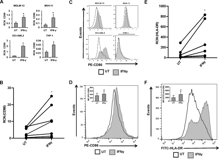
IFNγ promotes an M1-related phenotype in AML cells. AML cell lines MOLM-13, MV4-11, OCI-AML3, and THP-1 (n = 3 or more separate experiments each) and primary AML apheresis samples were treated with or without 10 ng/ml IFNγ for 18 h (qPCR) or for 24 h (flow cytometry, except for MOLM-13 treated for 48 h). A and B, CD86 expression in AML cell lines (A) and primary AML apheresis samples (B, n = 7 donors) was measured by qPCR. C and D, CD86 expression in AML cell lines (C) and primary AML apheresis samples (D, n = 7 donors, representative histogram shown; inset bar graph depicts all donors) was measured by flow cytometry. E and F, HLA-DR expression in primary cells was measured by qPCR (E, n = 6 donors) and flow cytometry (F, n = 7, representative histogram shown; inset bar graph depicts all donors). *, p ≤ 0.05. Error bars, S.D.

