FIGURE 1.
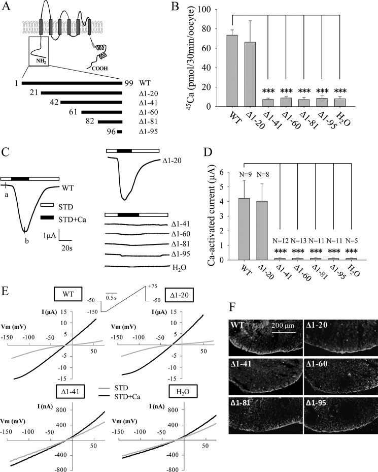
Channel function of human TRPP3 N-terminal truncation mutants. A, putative membrane topology predicted for TRPP3 (top) and five truncated mutants with indicated positions of the starting amino acid residue (bottom). B, radiolabeled 45Ca uptake in X. laevis oocytes expressing TRPP3 wild-type (WT) or mutants at day 3 following RNA injection. Oocytes injected with H2O were used as a negative control. Data were averaged from three independent experiments. *** indicates p < 0.001. C, representative whole-cell current traces obtained from Xenopus oocytes expressing TRPP3 WT or an indicated truncation mutant, using TEVC. Oocytes were voltage clamped at −50 mV. Data from H2O-injected oocytes served as a negative control. Currents were measured using the standard Na+-containing extracellular solution without (STD) or with (STD+Ca) 5 mm CaCl2. D, averaged Ca2+-activated currents obtained at −50 mV from oocytes expressing TRPP3 WT or an indicated mutant or those injected with H2O. Currents were averaged from three independent experiments with total numbers of tested oocytes, as indicated. *** indicates p < 0.001. E, representative current-voltage relationship curves obtained using a voltage ramp protocol, as indicated, before (STD) and after (STD+Ca) addition of 5 mm CaCl2 at the time point marked with a and b, respectively, in C. F, representative immunofluorescence data using oocyte slices, showing expression of TRPP3 WT and indicated truncation mutants.

