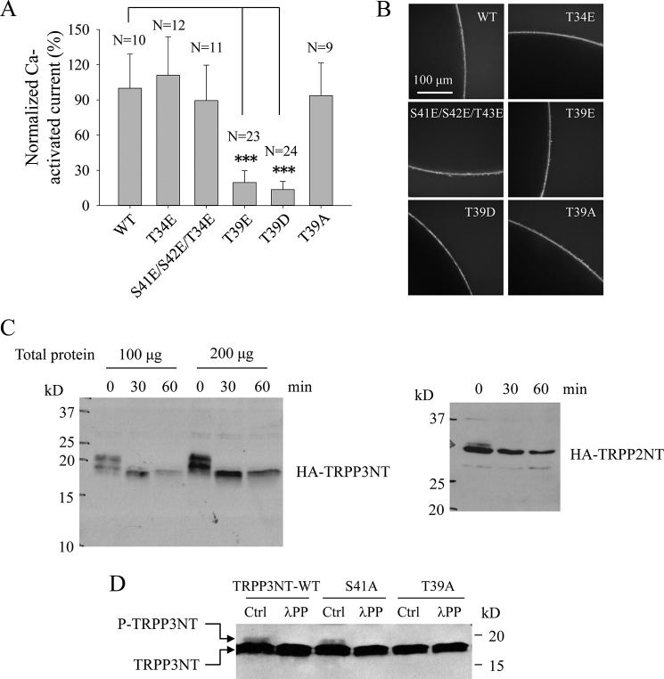
FIGURE 7.
Roles of Thr-39 and its phosphorylation in regulating TRPP3 channel function. A, averaged currents obtained from oocytes expressing TRPP3 WT, T34E, S41E/S42E/T43E (triple S41E, S42E, and T43E mutations) and T39E, T39D, or T39A. Currents at − 50 mV were averaged from three independent experiments with indicated total numbers of tested oocytes and normalized to that of TRPP3 WT. *** indicates p < 0.001. B, representative whole-mount immunofluorescence data showing the PM expression of TRPP3 WT or a mutant in oocytes. C, phosphorylation state of the TRPP3 N terminus assessed by λPP treatment. HA-tagged TRPP3NT and TRPP2NT (as a positive control) were transfected into HEK cells where cell lysates were treated with λPP for the indicated periods of time. Shown are representative blots from three independent experiments. D, phosphorylation of TRPP3NT WT and TRPP3NT containing the S41A or T39A mutation. These three constructs were transfected into HEK cells, and resulting cell lysates were treated without (Ctrl) or with λPP for 30 min. Shown are representative blots from three independent experiments.

