FIGURE 1.
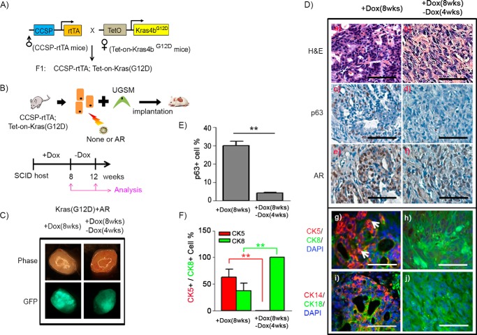
p63+ basal/progenitor cells possess differentiation potential to CK8+ luminal cells in Kras(G12D)+AR tumors. A, the CCSP-rtTA;Tet-on-Kras4bG12D mouse strain carrying doxycycline (Dox)-inducible Kras(G12D) was generated by crossing CCSP-rtTA mice with Tet-on-Kras4bG12D mice. B, prostate epithelial cells were isolated from prostate tissue of CCSP-rtTA;Tet-on-Kras4bG12D mice and transduced with AR along with GFP reporter by lentiviral infection. The infected cells were mixed with UGSM cells and implanted under the kidney capsule of CB.17SCID/SCID mice. The host mice were given drinking water containing Dox for 8 weeks to allow Kras(G12D) expression, followed by a Dox withdrawal period for an additional 4 weeks (a total of 12 weeks) to shut down Kras(G12D) expression. C, phase and fluorescence images of Kras(G12D)+AR grafts derived from 8 weeks of Dox induction (+Dox) or 8 weeks (wks) of Dox induction plus 4 weeks of Dox withdrawal (−Dox). Green fluorescence indicates that prostate cells were successfully infected by AR lentivirus. D, histological analysis of the regenerated grafts by H&E (a and b) and IHC staining of p63 (c and d), AR (e and f), CK5 (red)/CK8 (green)/DAPI (blue) (g and h), and CK14 (red)/CK18 (green)/DAPI (blue) (i and j). Scale bars = 50 μm. The white arrows indicate CK5+CK8+ cells. E and F, the number of p63+, CK5+, and CK8+ and total number of cells in tubules of regenerated tissue were counted. The percentage of p63+ (E) and CK5+ and CK8+ cells (F) per regenerated tubule was calculated. **, p < 0.01.

