FIGURE 5.
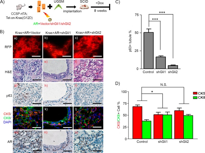
Expression of Gli1 and Gli2 facilitates proliferation of p63+ basal/progenitor cells in Kras(G12D)+AR tumors. A, schematic of the in vivo prostate regeneration assay, including lentiviral infection and schedule for Dox induction. Prostate epithelial cells derived from CCSP-rtTA;Tet-on-Kras4bG12D transgenic mice were transduced with AR and control vector, shRNA-Gli1, or shRNA-Gli2. The transduced cells were mixed with UGSM and implanted under the renal capsule. Host SCID mice were fed Dox in the drinking water for 8 weeks. B, RFP fluorescence (a–c), H&E (d–f), and IHC staining of p63 (g–i), CK5 (red)/CK8 (green)/DAPI (blue) (j–l), and AR (m–o) in regenerated tissues of Kras(G12D)+AR+Vector, Kras(G12D)+AR+shGli1, and Kras(G12D)+AR+shGli2. Scale bars = 50 μm. C and D, the number of p63+, CK5+, and CK8+ and total number of cells in tubules from Kras(G12D)+AR+vector and Kras(G12D)+AR+shGli1/2 regenerated tissues was counted. The percentage of each type of cells was calculated. The ratio of CK5 to CK8 cell number was significantly reduced in Kras(G12D)+AR+shGli1 but not Kras(G12D)+AR+shGli2 in comparison with Kras(G12D)+AR+Vector. Two-way ANOVA analysis was applied. *, p < 0.05; ***, p < 0.001; N.S., not significant.

