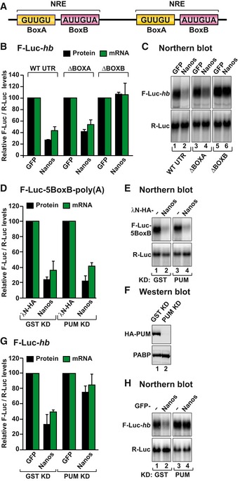
Figure 2. Dm Nanos exhibits intrinsic repressive activity.

- Schematic representation of the Nanos response element in the 3′ UTR of hb mRNA.
- The activity of GFP‐tagged Dm Nanos was tested in S2 cells expressing an F‐Luc reporter containing the hb 3′ UTR (either wild type or mutants lacking the BoxA or BoxB sequences). A plasmid expressing GFP served as a negative control. F‐Luc activity (black bars) and mRNA levels (green bars) were analyzed as described in Fig 1B. The panel shows mean values ± standard deviations from three independent experiments.
- Northern blot of representative RNA samples corresponding to the experiment shown in (B).
- Tethering assay using Dm Nanos and the F‐Luc‐5BoxB reporter in S2 cells depleted of PUM or control cells treated with a dsRNA targeting bacterial GST. A plasmid expressing R‐Luc mRNA served as a transfection control. The F‐Luc activity (black bars) and mRNA levels (green bars) were normalized to those of the R‐Luc transfection control and analyzed as described in Fig 1B. The panel shows mean values ± standard deviations from three independent experiments.
- Northern blot of representative RNA samples corresponding to the experiment shown in (D).
- Western blot analysis of S2 cells depleted of PUM and expressing HA‐PUM. Endogenous PABP served as a loading control.
- The ability of Nanos to repress the F‐Luc‐hb reporter was tested in S2 cells depleted of PUM as described in (D). The panel shows mean values ± standard deviations from three independent experiments.
- Northern blot of representative RNA samples corresponding to the experiment shown in (G).
Source data are available online for this figure.
