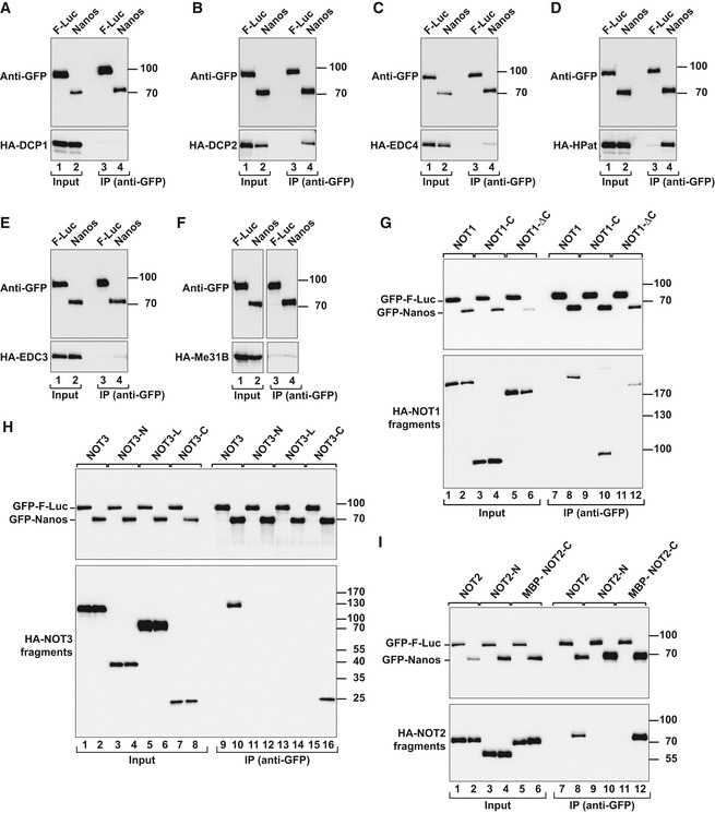
Figure EV2. Dm Nanos interacts with decapping factors and the NOT module.

-
A–FCo‐immunoprecipitation assays using GFP‐tagged Dm Nanos (full length) and HA‐tagged decapping factors. Samples were analyzed as described in Fig EV1G–M.
-
G–IWestern blot analysis showing the interaction of GFP‐tagged Dm Nanos and HA‐tagged NOT1, NOT2, and NOT3 (either full length or the indicated fragments). Proteins were immunoprecipitated from RNase A‐treated cell lysates using anti‐GFP antibodies. GFP‐F‐Luc served as a negative control. For the detection of GFP‐tagged proteins, 3% of the input and 10% of the bound fractions were analyzed by Western blotting. For the detection of HA‐tagged NOT1, 1.5% of the input and 35% of the bound fractions were analyzed, whereas for HA–NOT2 and HA–NOT3 proteins, 1% of the input and 30% of the immunoprecipitates were analyzed.
Source data are available online for this figure.
