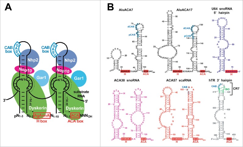
Figure 1.

Structure of H/ACA RNAs and RNPs. (A) Schematic structure of eukaryotic double hairpin H/ACA RNPs. The consensus sequences of the conserved H and ACA boxes are highlighted in red boxes. The consensus CAB box sequences are in blue. Usually, the target uridine (Ψ) selected for pseudouridylation is separated by 13–15 nucleotides from the H and ACA box. Arrangement of the associated H/ACA RNP proteins dyskerin, Gar1, Nop10 and Nhp2 is indicated. (B) Computer-predicted secondary structures of human H/ACA RNAs used in this study. The AluACA7, AluACA17 (black), U64 (blue), ACA30 (pink), ACA57 (red) and telomerase RNA (hTR, gray) sequences are indicated in different colors. The conserved H and ACA motifs are in red boxes. The CAB boxes of ACA57 and hTR and the distal and proximal CAB boxes (dCAB and pCAB) of AluACA RNAs are highlighted in blue circles.
