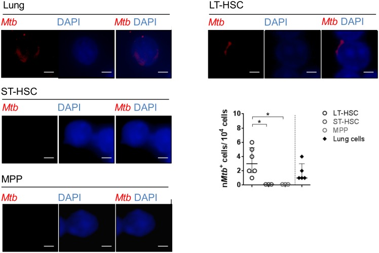
Fig 3. Detection of Mtb in cells of the lung and hematopoietic cells of Mtb-infected mice by histology.
Rhodamin-auramin stainings of representative LT-pHSCs (n = 6), ST-pHSCs (n = 3), MPPs (n = 3) as well as cells of the lung (n = 5) at day 28 p.i. For each sample (cell sort) 10,000 cells were screened per slide for Mtb positive cells. At least 3 images were taken from each slide of each sample. Rhodamin-auramin stainings were screened on high power (100×) and verified under oil immersion using a fluorescent microscope. Analyses were carried out using ProGres Capture Pro 2.8.8. (Mtb, red; nuclei, blue). Shown are representative data (cropping of images) for staining of LT-pHSCs, ST-pHSCs, MPPs and cells of the lung. scale bar: 10 μm. *P ˂ 0.05 by Mann-Whitney test.

