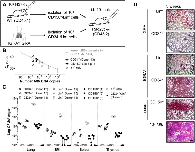
Fig 6. Intratracheal transfer of Mtb infected human and murine pHSCs leads to Mtb growth and increased cellularity into the lungs in transplanted hosts.
(A) Injection of Lin-CD34+ and Lin+ cells from blood of IGRA+ human donors (Donor 12–14) and mouse LT-pHSCs from bone marrow 28 days p.i. into the trachea of Rag2–/–Il2rg/–mice (3 mice/population). Transfer of 102 CFUs Mtb was used as positive (n = 3), uninfected pHSCs and Lin+ cells of an IGRA−donor (Donor 3; n = 1) as negative, control. Recipients were analyzed after 3 weeks. (B) Monitoring of Mtb infection by TaqMan PCR using probes that target MPB64 and IS6110 together on genomic DNA of 105 lung cells 3 weeks upon transfer. PCRs were performed in technical triplicates and normalized to murine GAPDH. (C) CFU Mtb growth on Middlebrook 7H11 agar in cells of lung, spleen, thymus and non-separated, 105 bone marrow cells 3 weeks upon transfer (n = 3/population). Shown are data from 3 independent experiments. (D) Histopathology of representative lung sections 3 weeks upon transfer. Lungs were stained with hematoxylin/eosin, screened with 5×objectives and verified using a light microscope. Shown are representative data from 3 independent experiments. Data are shown as median + interquartile. Scale bar: 100 μm.

