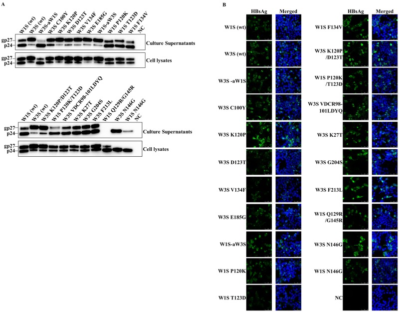
Fig 4. Detection of the wild-type and mutated HBsAg with Western blot and immunofluorescence analysis.
(A) Western blot analysis. The HEK 293-T cells were transfected with plasmid vectors expressing the wild-type and mutants of W3S and W1S HBsAg. The expressed HBsAg was purified from the culture supernatant and the cell lysate 72 h after transfection and subjected to Western blot with an anti-Xpress mAb against the Xpress tag to detect the HBsAg. The upper panels shows blots from the culture supernatants and the lower one blots from cell lysates. (B) Immunofluorescence assay. The cells on the culture slides were fixed 72 h after transfection and stained with an anti-Xpress mAb against the Xpress tag followed by an anti-mouse IgG conjugated with Alexa Fluor 488. The cell nuclei were stained with DAPI. Non-transfected cells were used as a negative control (NC). Each experiment was performed independently three times and one representative result is shown. wt: wild-type.

