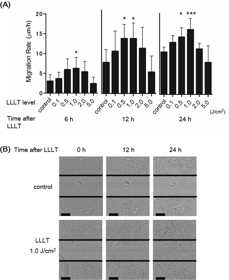
Fig 3. Effects of LLLT with a CO2 laser on HDF migration.
A cell migration assay of HDFs treated with LLLT under several irradiation powers (0.1, 0.5, 1.0, 2.0, or 5.0 J/cm2). The non-irradiated group served as the control. (A) Migration rates (0.1, 0.5, 1.0, 2.0, or 5.0 J/cm2) are expressed as migration distance/time (μm/h). The non-irradiated group served as the control. Results are expressed as the mean ± SD of three independent experiments. *p<0.05, ***p<0.001 compared with the non-irradiated group (Tukey-Kramer test). (B) Images show the wounded cell monolayers at 0, 12, and 24 h after wounding with non-irradiated or irradiated (1.0 J/cm2) HDFs. The line indicates the wound edge at the start of the experiment (0 h). Bar = 300 nm. The migration of irradiated HDFs was promoted compared with the control.

