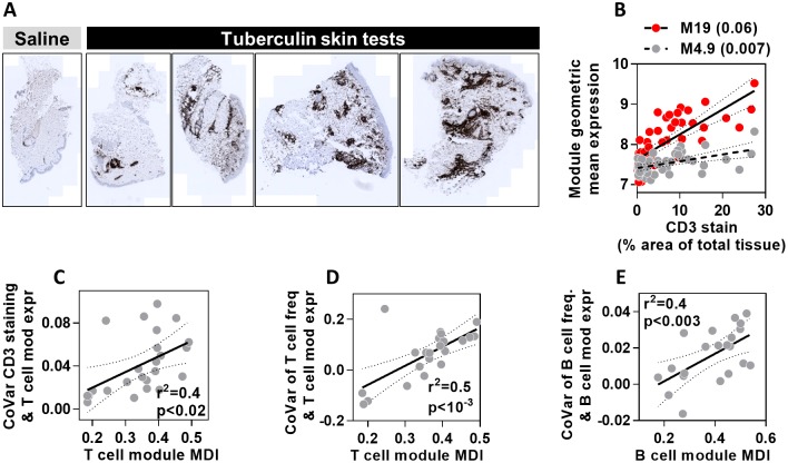
Fig 6. MDI scores predict covariance between module gene expression and cell frequency in skin and lymph nodes.
(A) Representative images of CD3 immunohistochemical staining in skin biopsies at the site of saline injection or tuberculin skin tests. (B) Relationship between geometric mean expression of two T cell modules which show highest (M19) and lowest (M4.9) covariance (shown in brackets) with quantitation of CD3 immunostaining. Each data point represents measurements derived from a TST in separate individuals. Regression lines and 95% confidence limits are shown for each module. (C) The relationship between covariance of CD3 immunostaining in TST biopsies and T cell module expression, with the MDI for all T cell modules. (D-E) The relationship between covariance of T or B cell frequency and cell-type specific module expression, with MDI for each module. Data points represent data derived from individual modules, giving regression lines with 95% confidence limits, Spearman rank correlation coefficients (r2) and p values.

