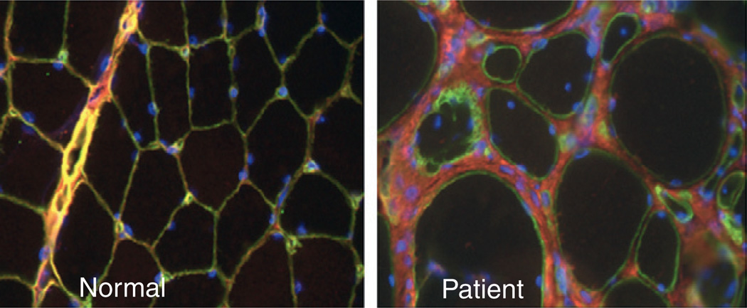
Figure 5.6.
Immunolocalization of collagen VI in the muscle of a patient with a dominant negative mutation in collagen VI. In the normal biopsy, collagen VI (red) overlaps with the basement membrane (green), resulting in a yellow color. In the patient’s biopsy there is a considerable amount of collagen VI immunoreactivity in the matrix; however, the colocalization with the basement membrane is lost, resulting in the green color of the basement membrane. Nuclear counterstain in blue.

