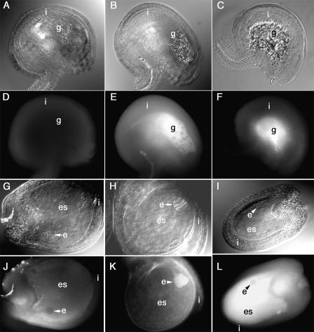
Figure 5.
Callose accumulated in the gametophytes and embryo sacs of aborting ovules. Using Nomarsky optics, the internal anatomy of ovules is shown in A to C and G to I, while D to F and J to L show callose fluorescence. The ovules in A, D, G, and J were from healthy plants, while those in B, C, E, F, and H to L were harvested from salt-stressed plants. A, Anatomy of a healthy ovule. Starch-containing plastids were present in the gametophyte (g). B, One day after plants were stressed, additional starch grains were produced in the gametophyte. C, Two days after plants were stressed, the central portion of the ovule, where the gametophyte had been, contained cellular debris. D, In healthy ovules, the integuments (i) synthesized only small amounts of callose. E and F, In stressed ovules, gametophyte cells synthesized copious amounts of callose. G, In healthy ovules, the embryo (e) developed within the embryo sac (es). J, Only minute amounts of callose were present in healthy embryo sacs. H and K, One day after plants were salt stressed, the embryos synthesized callose. I, One day later, embryo development ceased at the globular stage, and large amounts of callose were present in the embryo, embryo sac, and endothelial walls (L).

